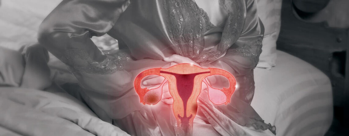
Tumors of female genital organs can be benign or malignant, both external and internal reproductive organs can suffer. The most common forms of female cancer are cervical cancer, endometrial cancer, ovarian cancer, vulvar cancer. Uterine sarcomas and gestational trophoblastic disease are less common. Female genital organs tumors are serious medical and social problem, therefore they require early detection and high-quality comprehensive treatment.

If you have been diagnosed with a malignant neoplasm of uterus, cervix, ovaries or other organs of reproductive system – do not delay! Contact the specialists of Oncology Department of Maimonides Multidisciplinary Medical Center for help. Our clinic works in close cooperation with the best Israeli oncology centers, we chose Israeli medicine as a model for providing medical services because it is one of the best in the world Each individual case of female reproductive system malignant disease is jointly managed by attending oncologyst, the head of the department – Dr. Iryna Stefanska and responsible oncologist directly from Israel.
The Maimonides Medical Center works under the patronage “Keren Or for our Child” the Charitable Foundation, so every patient who needs expensive treatment can receive financial assistance for frequent or all medical and diagnostic manipulations. Fund assets are replenished by caring people and various companies.
Types of female reproductive system tumors
There are many types of oncological lesions of female genital organs, including both benign and malignant. Tumors can occur in all external and internal reproductive organs, can be single or multiple, primary and metastatic. Let's consider the most common tumors.
1. Cervical cancer is usually squamous. Adenocarcinoma is rarely diagnosed. Human papillomavirus infection is the cause of most cervical cancer types. Cervical neoplasia is often asymptomatic.
The first symptoms of cervical cancer are usually irregular, often it can be postcoital (after sex) bleeding. Diagnosis includes Pap test and biopsy. If possible, disease clinical stage is determined in combination with imaging and pathological examination results. For early-stage disease, treatment usually involves surgical resection or radiation therapy, along with chemotherapy if the disease is locally advanced. If cancer has metastasized far away, chemotherapy is often used as only treatment.
Cervical cancer is the 3rd most common gynecological cancer among women. The average age at the time of diagnosis is 50 years.
2. Vulvar cancer (malignant lesion of female external genitalia) is usually a squamous cell carcinoma. It is most common among elderly women. It usually appears as a palpable tumor. Diagnosis is based on biopsy data.
Treatment usually involves tumor surgical removal. Vulvar cancer is the 4th most common cancer in women. The average age of diagnosis is about 70 years, disease frequency increases with age. Cancer of vulva often occurs on labia majora. It rarely affects labia minora, clitoris or vaginal glands. Tumor usually develops slowly over several years.
3. Vaginal cancer, is usually a squamous cell carcinoma and most often develops in women over 60 years age. The most common symptom is vaginal bleeding. Diagnosis is based on biopsy data.
Tumor Surgical removal and radiation therapy are used to treat most cases. Primary vaginal cancer is rare. Vaginal metastases or local spread from nearby gynecological anatomical structures are more common than primary tumors of this location. The average age of diagnosis is 60-65 years.
4. Endometrial cancer, is usually represented by adenocarcinomaThe disease most often manifests itself in form of postmenopausal uterine bleeding. Diagnosis is based on biopsy data. The stage of disease is determined during surgery.
Treatment includes uterus with appendages removal, and for patients with recurrence high risk also pelvic and para-aortic lymph nodes excision. In the later cancer stages, radiation therapy, hormone therapy, or chemotherapy are usually prescribed. Endometrial cancer is most common in high-income countries with high obesity rates. This type of malignant disease is on the 4th place among the most common types of cancer in women. Endometrial cancer most often develops mainly in women in postmenopausal period. The average age of patients at the time of diagnosis is 61 years. Most cases are diagnosed in women aged 45 to 74 years.
5. Uterine sarcomas are a group of heterogeneous cancerous tumors of high malignancy degree that develop in uterus body. Typical manifestations include abnormal uterine bleeding and pelvic pain or palpable mass presence. If uterine sarcoma is suspected, endometrial biopsy or dilation and curettage can be performed, but results are often false negative. Most sarcomas are diagnosed histologically after hysterectomy or myomectomy. Treatment requires total uterus removal with appendages. Chemotherapy and radiation therapy are indicated for widespread process.
6. Ovarian cancer often has difficult prognosis because it is usually diagnosed in late stages. Symptoms are usually absent or nonspecific. Diagnostic methods usually include ultrasonography, CT or MRI and determination of tumor markers (for example, cancer antigen CA 125). Diagnosis is established based on histological examination. The stage of disease is determined during surgery.
Treatment includes uterus removal with appendages and, usually, chemotherapy. Ovarian cancer is the second most common gynecological cancer. In developed countries, incidence rates are higher.
7. Gestational trophoblastic disease (alveolar drift, chorionic carcinoma) is proliferation of trophoblast tissues in pregnant women or women who have recently ended pregnancy. Symptoms may include excessive uterine enlargement, vomiting, vaginal bleeding, and preeclampsia, which usually occurs on early terms of pregnancy.
Diagnosis includes amout of hCG (human chorionic gonadotropin) beta subunit measuring, pelvic ultrasonography and biopsy to confirm the diagnosis. Tumors are removed using vacuum curettage. If disease persists after tumor removal, chemotherapy is prescribed. Gestational trophoblastic disease develops during or after uterine or ectopic pregnancy. Risk increases during pregnancy in women in the later stages of reproductive life, especially after the age of 45. During pregnancy, disease usually leads to spontaneous abortion, eclampsia or fetus death.

Causes and symptoms of female genital organs tumors
Among the risk factors for female reproductive system malignant neoplasms development should be mentioned:
- Infection caused by human papillomavirus (HPV).
- Cervical intraepithelial neoplasia.
- Increased risk of contracting sexually transmitted diseases (for example, early age of sexual life onset or early first birth, multiple sexual partners, sexual partners at high infection risk presence).
- History of vulvar or vaginal squamous intraepithelial neoplasia or cancer.
- Anal intraepithelial neoplasia or cancer.
- Oral contraceptives application
- Smoking.
- Immunodeficiency.
- History of ovarian cancer in relative of the 1st degree consanguinity.
- Childbirth absence in anamnesis.
- First late childbirth.
- Early menstruation onset.
- Late menopause.
- Personal or family history of endometrial, breast, or colon cancer.
- Hyperestrogenia (high levels of estrogen circulating and no or low progesterone levels).
- Age over 45 years.
- Obesity.
- Application of tamoxifen for > 2 years.
- Lynch syndrome.
- Radiotherapy in pelvic organs area in the anamnesis.
Рак шийки матки на ранніх стадіях може протікати безсимптомно. Коли з’являються симптоми, вони зазвичай включають нерегулярні вагінальні кровотечі, які можуть бути посткоїтальними або іноді спонтанно траплятися між менструаціями. Більші пухлини частіше проявляються спонтанними кровотечами, а також можуть викликати виділення з неприємним запахом або біль.
При поширенні раку може статися обструкція сечовивідних шляхів, з’явитися болі в спині, набряк нижніх кінцівок через венозну або лімфатичну обструкцію.
У більшості пацієнтів з раком піхви спостерігається аномальна міжменструальна піхвова кровотеча, часто постменопаузальна або посткоїтальна. У деяких пацієнток також спостерігаються рідкі вагінальні виділення або диспареунія (біль при статевому акті). У деяких пацієнток відзначається безсимптомний перебіг захворювання.
Більшість сарком тіла матки проявляються патологічними матковими кровотечами, рідше, тазовими болями, відчуттям заповненості живота, появою пухлин піхви, частим сечовипусканням або тазовими пухлинами, що пальпуються.
Рак яєчників може протікати тривалий час безсимптомно. Симптоми, у разі їх наявності, неспецифічні (наприклад, диспепсія, здуття живота, раннє насичення під час їжі, зміна частоти та характеру випорожнень, прискорене сечовипускання). Потім, як правило, з’являються тазові болі, анемія, виснаження та збільшення живота за рахунок збільшення яєчника або асциту. Герміногенні або стромальні пухлини, які продукують гормони, можуть виявлятися функціональними порушеннями (наприклад, гіпертиреоз, вірилізація).
Більшість жінок з раком ендометрію мають аномальні маткові кровотечі (наприклад, постменопаузальна кровотеча, пременопаузальна міжменструальна кровотеча, овуляторна дисфункція).

Методи діагностики пухлин жіночих статевих органів
Як відомо, чим раніше встановлений діагноз злоякісної пухлини, тим кращий прогноз та результат лікування. Тому якісна та швидка діагностика – це надзвичайно важливий етап. В клініці Маймонідес використовується лише сучасне обладнання експертного класу, всі наші лікарі досконало володіють всіма методиками обстеження пацієнтки з підозрою на злоякісні новоутворення репродуктивної системи, уміють безпомилково інтерпретувати отримані дані, що допомагає їм у створення сучасного, індивідуального та ефективного плану лікування.
План діагностичного пошуку залежить від підозри лікаря на конкретне захворювання. Як правило, використовується комбінація х наступних діагностичних методів:
- Цитологічна діагностика – рідинна цитологія, ПАП-тест.
- Colposcopy – огляд шийки матки зі збільшенням з допомогою сучасного приладу – кольпоскопу.
- Ultrasound органів малого тазу.
- Neoplasm biopsy з подальшим патогістологічним та молекулярно-генетичним дослідженням.
- КТ, МРТ з контрастом ОМТ, ОГК, ОЧП або інших органів та тканин при підозрі на віддалені метастази. Остеосцинтиграфія, ПЕТ-КТ, ПЕТ-МРТ для пошуку віддалених метастазів.
- Пухлинні маркери в крові – бета-ХГЛ при хоріонкарциномі або раковий антиген СА 125 при раку яєчників.
After biopsy we send all histological materials to the world's best pathohistological laboratories (Israel, Germany, USA).Thanks to such double checks, we are absolutely sure of diagnosis correctness and effectiveness of the selected treatment tactics.
Innovative molecular genetic tests to our patients after biopsy. . This is a mandatory part of modern diagnosis of oncological diseases. Thanks to molecular genetic diagnostics, we can choose the most effective treatment regimens, because the response to certain drugs action depends on the type of malignant changes.
An example of such modern diagnostics are test systems for molecular genetic testing. Such as the Foundation One and Caris Molecular Testing. These tests make it possible to determine certain types of mutations in tumor cells, as well as certain receptors presence on the surface of these parasitic cells. These data are subsequently used to create individual immunobiological therapy drugs or to select target therapy drugs. In modern medicine this approach is called personalized oncology. Our patients have access to all this benefits of this innovative treatment.

Сучасне лікування пухлин жіночих статевих органів
Однією з найважливіших переваг лікування в МЦ Маймонідес є застосування комплексного підходу до кожного випадку злоякісного захворювання. В боротьбі з пухлиною лікар застосовує увесь доступний арсенал методів. Терапія завжди є комбінацією з двох, трьох, а то й більше методик.
Відділення онкогінекології надає доступ своїм пацієнткам до найсучасніших методів лікування пухлин репродуктивних органів.
На базі ізраїльських клінік-партнерів медичного центру Maimonides постійно проходять клінічні дослідження з підтвердження ефективності інноваційних препаратів та методів терапії різноманітних новоутворень. Тому всі наші пацієнтки при бажанні можуть брати участь в цих експериментальних дослідженнях абсолютно безкоштовно.
As a rule, patients are selected for research in case when standard treatment scheme was inefficient, and participation in research is the only real chance for a healthy future.
Одним з основних методів лікування злоякісних пухлин жіночих статевих органів є хірургічна операція. Всі операції, які виконують наші хірурги спрямовані на повне одужання пацієнтки одночасно зі збереженням її репродуктивної функції. В основному всі операції вдається виконувати малоінвазивними методиками – лапароскопічно та гістероскопічно. До відкритої хірургії з видаленням матки чи додатків вдаються лише в крайніх випадках.
Для наших пацієнток доступні інноваційні хірургічні методики лікування пухлин репродуктивних органів:
- Роботизована гінекологічна хірургія з допомогою установки «Да Вінчі».
- Роботизована операція з допомогою установки Single Port.
- Однопортова відеоторакоскопія в онкогінекології.
- Хірургічна циторедукція в комбінації з хіміотерапією.
- Фотодинамічна терапія при захворюваннях шийки матки.
- Евісцерація органів малого тазу.
Збереження фертильності у молодих жінок з онкогінекологічними захворюваннями – це основний пріоритет нашого відділення.
Наші спеціалісти в області гінекології та репродуктивної медицини пропонують пацієнткам перед початком радикального лікування a delayed motherhood program before starting radical treatment. У разі згоди репродуктологи проводять стимуляцію суперовуляції, пункцію та забір яйцеклітин з їх подальшою кріоконсервацією.
У випадку, якщо у жінки вже є чоловік, то може проводитись процедура екстракорпорального запліднення та кріоконсервація уже готових ембріонів. Після отриманого комплексного лікування онкопатології навіть у випадку втрати фертильності пацієнтка або подружня пара зможуть в майбутньому мати своїх генетично рідних дітей шляхом виконання процедури ЕКО.
Chemotherapy раку репродуктивних органів проводиться, як правило, на другому етапі лікування після радикального хірургічного втручання. Основна мета хіміотерапії – боротьба з метастазами та всіх злоякісних клітин, щоб уникнути рецидиву хвороби. Кількість курсів хіміотерапії для пацієнток із раком яєчників, ендометрію на ранніх стадіях становить від 3 до 5. Якщо виявлено рак 3-4 стадій, необхідно більше 6 курсів введення препаратів.
Лікарі онкологи у лікуванні раку яєчників використовують один із найефективніших сучасних методів хіміотерапії – інтраперитонеальну або внутрішньочеревну хіміотерапію (ІР та НІПЕС). Дана методика передбачає введення висококонцентрованого розчину препаратів безпосередньо в черевну порожнину, за допомогою спеціального пристрою, що імплантується. Планомірна взаємодія ліків з органами малого тазу та черевної порожнини дозволяє досягти знищення максимальної кількості ракових клітин.
Radiation therapy у лікуванні раку репродуктивної системи у жінок показана у разі неможливості хірургічного втручання (на пізніх стадіях раку) для полегшення симптомів перебігу хвороби, а також у післяопераційному періоді для запобігання рецидивам.
Радіоонкологи нашої клініки використовують сучасні види радіотерапії, зокрема променеве лікування з регульованою інтенсивністю опромінення (IMRT). Даний метод лікування раку передбачає точковий вплив на новоутворення та збереження здорових органів та тканин. Тривалість та обсяги опромінення за методом IMRT значно менші, ніж при стандартній радіотерапії.
Лікування раку на пізніх стадіях, згідно з сучасними протоколами, передбачає використання цільової чи targeted therapy. Лікарі клініки Маймонідес застосовують новітні біопрепарати, у складі яких містяться моноклональні антитіла. Дані антитіла впливають безпосередньо на злоякісні клітини новоутворення, зупиняють їх ріст та поділ. Здорові тканини при цьому страждають, що відрізняє принципово таргетну терапію від класичної хіміотерапії.
Гормональна терапія активно використовується в комплексній терапії злоякісних новоутворень жіночих статевих органів. Гормонотерапія дозволяє скоротити вироблення гормонів, необхідних для росту пухлини, та домогтися зупинки метастазування, зменшення новоутворення у розмірах. Гормонотерапія може бути використана як самостійний метод лікування при раку яєчників пізніх стадій, а також може бути призначена замість хіміотерапії на другому етапі лікування на I-II стадіях.
У кожному окремому випадку рішення про комбінацію тих чи інших методик лікування приймається спільно командою спеціалістів. Кожна жінка та її хвороба відрізняються, тому наші лікарі, спираючись на свій досвід в лікуванні онкогінекологічних патологій, часто виходять за рамки стандартних протоколів, змінюючи схеми лікування та дози необхідних препаратів, методику опромінення так, щоб отримати якнайкращі результати для своїх пацієнтів.

