The group of skin oncological diseases includes benign, malignant and borderline neoplasms. Malignant skin tumors are the most common type of cancer, and in most cases, they develop on skin areas that are exposed to insolation (sun exposure).The highest prevalence is among outdoor workers, athletes, and sunbathers. Patients with light skin are most prone to cancer development. Malignant skin tumors can develop years after radiation therapy or carcinogens exposure .
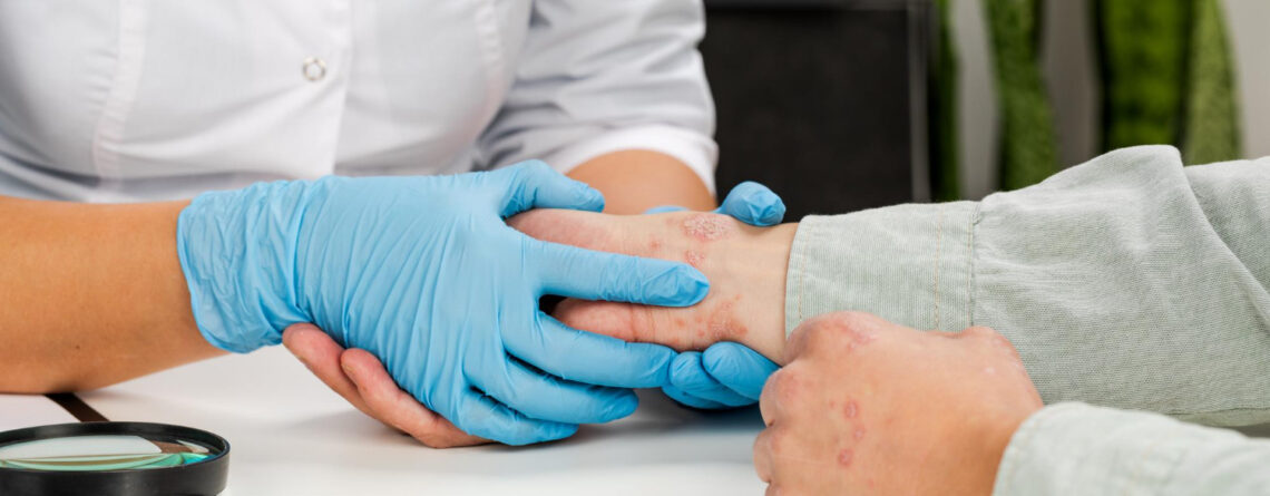
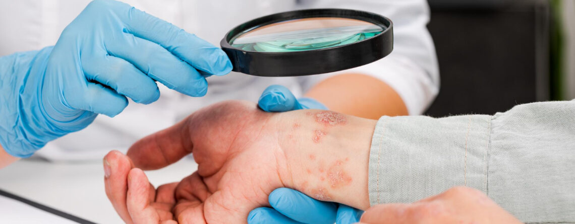
If you notice a suspicious neoplasm on your skin, you need to see a dermatologist for an examination. Doctor will carefully examine the neoplasm, the entire skin surface, and may prescribe additional examinations. If, during the diagnostic process, specialist suspects or diagnoses skin malignant neoplasm, patient must be referred to a qualified oncologist for consultation.

Such specialists work in Oncology Department of Maimonides Multidisciplinary Medical Center. Our clinic works not only according to Israeli medicine model, as one of the best in the world, but also in close and fruitful cooperation with well-known Israeli oncology centers. Each individual clinical case is jointly managed by the attending oncologist, the head of the department – Dr. Iryna Stefanska and responsible oncologist directly from Israel. Thanks to such work of a professional team, each patient receives high-quality and modern medical services, which significantly increases the chances of a full recovery.
Maimonides Medical Center works under the patronage of “Keren Or for our Child” the Charitable Foundation, , so our patients can count on financial assistance. Depending on situation, the fund can partially or even fully pay for necessary medical services for seriously ill patients.
One of the most important treatment advantages at Maimonides Medical Center is an individual approach to each patient. Of course, in their work, doctors are guided only by modern clinical recommendations and world treatment protocols, but they never use one examination template or treatment scheme for each patient, because there are no two identical people and absolutely similar diseases. Making up a treatment and diagnostic program, our specialists must take into account all factors: age, patient’s gender, main diagnosis, tumor features (histological and molecular genetic), the stage of oncological process, serious concomitant diseases presence and patient’s wishes regarding therapy process.
Treatment of any oncological diseases, especially malignant ones, is a complex process that requires several narrow specialists involvement at the same time. In our Oncology Department, based on the best Israeli clinics example, not only oncologists work, but a whole multidisciplinary team – oncologists, dermatologists, therapists, surgeons, radiation therapists, chemotherapists, plastic surgeons, rehabilitation specialists, psychologists and others . This ensures continuity of medical care for any patient, that is, all stages from diagnosis to complex treatment and recovery after it can be obtained within the walls of one medical institution.
Our department is equipped only with modern medical and diagnostic equipment of an expert class. If a certain narrow specialist or rare medical and diagnostic equipment is not available, patient is referred to subsidiary institutions of Maimonides Medical Center or our Israeli partner clinics, where he is guaranteed to receive the full range of necessary medical services, where he is guaranteed to receive the full range of necessary medical services.

Types of oncological skin diseases
All skin neoplasms can be divided into:
- benign,
- malignant,
- borderline (borderline).
Skin malignant neoplasms in adults are quite common. Among them, basal cell carcinoma (BCC), squamous cell carcinoma, and melanoma are the most common.
Benign skin neoplasms in one form or another are present in every person without exaggeration. These include nevi, fibromas, dermatofibromas, lipomas, hemangiomas, papillomas, and others. These neoplasms almost do not pose a danger to life, although they can sometimes become malignant (degenerate into malignant ones). Therefore, it is better to remove such neoplasms with modern minimally invasive techniques, for example, by cryodestruction or radio-wave coagulation.
Borderline neoplasms refer to precancerous (background) conditions, that is, at any moment such a tumor can turn into a malignant one and acquire the ability to metastasize. Most common are pigmented xerodermas and keratoacanthomas.
Less common forms of skin cancer are:
- Paget's disease (breast nipple cancer) or extramammary Paget's cancer (usually near the anus).
- Kaposi's sarcoma.
- Merkel cell carcinoma.
- Atypical fibroxanthoma.
- Tumors of skin appendages, such as sweat glands.
- Cutaneous T-cell lymphoma (mycosis fungoides).
- Bowen's disease – superficial squamous cell carcinoma.
- Keratoacanthoma can be a highly differentiated form of squamous cell carcinoma.
Basal cell carcinoma is the most common type of skin cancer. As a rule, basal cell carcinoma develops in people with fair skin and a sun exposure history, and very rarely in patients with dark, pigmented skin. Basal cell carcinomas are also associated with genetic syndromes. Basal tumors grow from cells near basal layer of skin. Metastases occur rarely, but local growth can be accompanied by pronounced destruction. Diagnosis is made based on biopsy data . Treatment depends on tumor characteristics and may include curettage and electrocoagulation, tumor surgical removal, cryodestruction, chemotherapy using topical chemotherapy, and sometimes radiation or chemotherapy.
Squamous cell carcinoma is the second most common form of skin cancer after basal cell carcinoma. Squamous cell carcinoma can develop on unchanged skin background, or on pre-existing keratosis background, in plaque area with oral cavity leukoplakia or in a post-burn scar. This is epidermal cells malignant tumorspreading to dermis. This type of skin cancer usually appears on open skin areas. Local destruction can be very pronounced and metastases can occur in the later stages. Diagnosis is made based on biopsy data Diagnosis is made based on biopsy data. Treatment depends on tumor characteristics and can be applied using curettage and electrocoagulation, surgical excision, cryodestruction or radiation therapy.
Melanomas occur mainly on skin, but can also develop on mucous membranes of oral cavity, in genital area, perineum and conjunctiva. Melanomas can also develop in eye choroid and in nail bed. It is the third most common type of skin cancer. Melanomas are various in size, shape, color (usually the tumors are pigmented) and in their propensity for invasive growth and metastasis. Melanoma arises from melanocytes – cells that contain pigment. With widespread metastases, the prognosis is unfavorable. The diagnosis is made based on biopsy data. Treatment is complex.

Skin cancer symptoms
Skin oncological lesions, unlike other tumor diseases, are very easy to detect in the early stages. To do this, you only need to monitor your skin and consult a dermatologist in time if a suspicious neoplasm appears on it.
Signs you need to pay attention, observing your skin:
- New elements on the skin appearance – nevi, nodules, various rashes, ulcers, pigmented or depigmented spots.
- Change in size, shape, mole structure, appearanceof non-uniformity of its color, signs of inflammation appearance around it or a depigmented rim, removal of blood, pus or blood from the nevus.
- Pain, burning or itching in neoplasm area on skin.
- frequent traumatization of a certain element on skin.
Diagnostic Methods of skin tumors
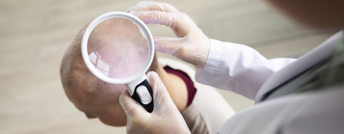
As a rule, diagnosis of skin neoplasms does not cause difficulties . If a suspicious element is detected on skin, you should consult a dermatologist. Specialist will conduct an examination , dermatoscopy, (an examination of element using a special dermatoscope device that magnifies image in 20 times), will summarize and systematize patient's complaints , collect medical and life anamnesis . If, according to initial examination data and dermatoscopy, neoplasm will be classified as borderline or suspicious for malignancy, patient will be offered a biopsy and sent to an oncologist.
Neoplasm biopsy is the main diagnostic method of skin malignant neoplasms. Technically, it is not difficult to perform a biopsy of skin neoplasms, because all elements are superficial. After biopsy we send all materials for revision to the best pathogistological laboratories in the world (USA, Israel, Germany). Thanks to such double checks, we are absolutely sure that diagnosis is made correctly and therapy is selected accordingly.
In addition to standard pathogistological examination, in the case of neoplasm malignant nature confirmation (especially often in case of melanoma), material is also examined by modern immunohistochemical, molecular genetic methods. This is necessary in order to determine certain types of mutations presence in malignant cells, certain receptors on their surface. In future, they become a target for innovative drugs created individually for each patient (targeted therapy, immunobiological drugs). Such test system for molecular genetic testing are the Foundation One and Caris Molecular Testing.
The second diagnosis step is disease stage establishing. That is, specialists assess the prevalence of malignant disease, regional and distant metastases presence. For this purpose, modern medical imaging techniques are used – lMRI, CT with or without contrast, ultrasound, PET-CT, PET-MRT, bone scintigraphy.
Based on received data, doctor formulates a detailed diagnosis indicating the stage of oncological process. Depending on diagnosis, effective treatment is selected for each patient. All necessary diagnostic procedures can be performed directly in clinic, without leaving Ukraine.

Modern treatment of skin tumors
All treatment procedures depend on the main diagnosis and the stage of oncological process. Benign or borderline neoplasms are treated only surgically, they do not require any other treatment. As a rule, neoplasms are removed by narrow excision without removing a part of healthy skin or by modern minimally invasive methods – electrocoagulation, cryodestruction, laser coagulation, radio wave ablation . After such manipulations, a scar almost never remains on skin, and after a certain time there will be no trace of tumor.
Electrocoagulation is neoplasm removal with a high-intensity current, widely used in oncodermatology to remove various benign skin tumors. This technique allows us to remove neoplasm quickly (in one visit) and absolutely painlessly. A specialist who knows all the techniques an electrocoagulator application is an Israeli dermatologist – Dr. Tomas. He removes all skin neoplasms using this modern technique: viral papillomas, senile keratomas, condylomas, atheroma, warts, molluscum contagiosum, hemangiomas, postacne, telangiectasia, rosacea, pigment nevi.
If, after diagnostic program, patient is diagnosed with skin malignant neoplasm, then treatment program is definitely expanded. If tumor is in early stages of its development and metastases are not detected, then the basis of treatment is tumor surgical removal by wide excision (within healthy tissues). Malignant neoplasms are not recommended to be removed by minimally invasive methods , but only by wide surgical excision. This prevents disease relapse. Often, after such interventions, a significant skin defect is formed, therefore either immediately or the second stage of surgical intervention is reconstructive and plastic surgery, especially if tumor was localized on face or another open body area.
If the disease is diagnosed at the 3-4 stage of oncological process, then treatment program must be supplemented with other methods - radiation therapy, chemotherapy, targeted and immunobiological therapy, etc.
Radiation therapy Radiation therapy is used in complex treatment after surgical intervention. As a rule, remote radiation using modern linear accelerators is applied. Modern equipment used at Maimonides Medical Center allows to perform targeted tumor radiation, minimizing dose to remaining healthy tissue. Modern linear accelerators available to our patients allow us to perform intensity modulated radiation therapy (IMRT), rotational (moving radiation) with beam intensity modulation (VMAT), as well as stereotaxic radiation therapy (SRT) and stereotaxic radiosurgery (SRS). Such equipment allows us to fight effectively with those cancer diseases that were previously considered incurable. Contact radiation therapy (brachytherapy) is also used for skin tumors, when radiation source is placed directly in tumor tissue.
Chemotherapy is also used in complex treatment of malignant skin neoplasms advanced stages. Unfortunately, such tumor as melanoma is resistant to standard chemotherapy (both systemic and contact). Therefore, in such patients, treatment is supplemented with modern method – targeted drugs, immunobiological drugs. Modern protocols of systemic chemotherapy include a combination of the following drugs: cisplatin, doxorubicin, methotrexate, bleomycin, etapozide, vismodegib.
Immunotherapy using anti-programmed cell death receptor 1 (PD-1) antibodies –pembrolizumab and nivolumab– prolongs survival in melanoma patients. Ipilimumab (monoclonal antibody to cytotoxic T-lymphocyte-associated antigen 4 [CTLA-4]) is another form of immunotherapy that may also increase life expectancy of patients.
Molecularly targeted therapy includes vemurafenib, dobrafenib, and encorafenibapplication, which work by inhibiting BRAFgene, resulting in slowing or stopping tumor cells proliferation. These drugs prolong life expectancy of patients with metastases.
A new TIL treatment protocol using "melanoma killers" is available for melanoma patients. TIL (tumour-infiltratinglymphocytes) protocol using "melanoma killers" is an innovative skin cancer treatment program used in Israel. It is effective even in metastatic cancer. This is a type cancer treatment biological method. From a distant metastasis, doctors obtain TIL cells and samples of abnormal cells. Healthy TIL cells are multiplied in laboratory and injected back into patient's body, where their number increases and ensures active fight of patient's immune system against cancer.
For each patient, the decision on certain treatment methods combination is made jointly by a team of specialists . Every person and their disease is different, so our doctors, relying on their experience in oncological pathologies treatment, often go beyond the standard protocols, changing treatment regimens and doses of necessary drugs, radiation method in order to get the best possible results for their patients.
