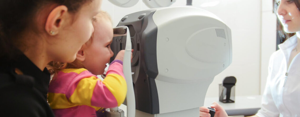
According to global statistical data, tumor lesions of eye occur in 4-5% of children, while they make up 4.3% of all neoplasms in childhood. If tumor is diagnosed immediately after the baby is born, it is considered congenital and is often caused by a mutation in child’s certain gene. Also, eye neoplasms can be acquired and appear already after child’s birth, regardless of age.
Eye tumors can be benign or malignant, eyelids, conjunctiva, iris, retina, optic nerve, orbital tissues can be affected. Tumors can be intraocular (growing from eyeball structures) and orbital (growing from anatomical structures of eye socket or eye auxiliary apparatus – eyelids, lacrimal glands, etc.). Most intraocular lesions are malignant in nature. Among such aggressive tumors in children, retinoblastoma prevails.

Eye tumors are dangerous not only for loss of vision, but also for life. That's why they are often malignant, have an aggressive, rapidly progressive course, early metastasize to distant organs, and grow into neighboring anatomical structures. Therefore, if such a disease is suspected or detected, child is referred for detailed diagnosis and comprehensive treatment in conditions of a modern Oncology Department.
Of course, treatment process of malignant disease is expensive, and this is no secret to anyone. Therefore, we have created conditions for our patients to receive financial assistance, which can cover all or part of treatment. Maimonides Medical Center works under the patronage of “Keren Or for our Child” the Charitable Foundation, which helps children to recover faster from a serious illness and lead a full future life.
This is exactly what Pediatric Oncology Department of Maimonides Multidisciplinary Medical Center is. It works under close supervision and in cooperation with the best Israeli medical oncology centers and specialists.If your child is suspected of having an eye neoplasm and you don't know where to start your journey to save child's life, feel free to contact us. During face-to-face meeting, doctor will explain all the details, talk about modern treatment possibilities and draw up an initial plan for follow-up examinations.
Children's oncology project in the field of eye neoplasms at Maimonides Medical Center is managed by the Israeli specialist Ido Didi Fabian. Together with our head of department, Dr. Yury Tymoshchuk, he supervises all clinical cases directly from Israel. This approach is unique, because in Ukrainian conditions, every small patient can get access to modern and effective Israeli medicine.
All our doctors are highly qualified and have extensive practical experience in helping children with cancer. Most of them have repeatedly trained on basis of the world's leading oncology centers. In their medical activities, specialists of Maimonides Medical Center are guided only by modern global treatment protocols, and not by domestic recommendations that are 20 years old. At the same time, the approach to each child is individual, treatment regimens are not templated for everyone, they are modified depending on child’s age, his main disease, concomitant pathology presence, child’s wishes or his parents.
Thanks to combination of such a patient-oriented approach, modern treatment protocols, innovative diagnostic and treatment equipment, highly qualified doctors, we achieve significant success in all eye tumors treatment, including the most dangerous childhood retinoblastoma.
Eye tumors are a very complicated pathology, especially malignant neoplasms. To receive quality medical care, one oncologist is not enough. Therefore, a whole multidisciplinary team works in our Pediatric Oncology Department. Oncologists, ophthalmologists, neurologists, neurosurgeons, plastic surgeons, pediatricians, psychologists, rehabilitation specialists, radiation therapists, diagnosticians and chemotherapists provide assistance to patient. If required narrow specialist or specific innovative diagnostic or treatment equipment is not available, patient, together with parents or custodians, is referred to subsidiary medical centers or Israeli partner clinics of Maimonides MC.

Symptoms of eye tumors in children
Manifestations of eye tumors primarily depend on the type of lesion. For example, a benign eyelid hemangioma will be visible to naked eye. It is more difficult to establish a timely diagnosis of intraocular malignant tumors, for example, retinoblastoma. A specific symptom in this case is white (milky) shade of pupil in affected eye. It is best seen in a photo with a flash – "cat's eye effect" or a white pupillary reflex – leukocoria. Normally, we see red-eye effect in such photos. This is the most common early symptom of retinoblastoma, which may be noticed by child's parents or by pediatrician or pediatric ophthalmologist during preventive examination.
Other symptoms of eye tumors:
- appearance of a squint in affected eye (strabismus),
- protrusion of one eye from orbit,
- change in usual color of iris in eye (color of affected eye in child gradually changes, becomes heterogeneous),
- various vision disorders – decreased visual acuity, limitation of peripheral vision, various photopsias (flies, stars in front of eyes), scotomas appearance – loss of a certain area from vision field (black spots appearance at the site of lesion),
- redness and pathological discharge from eye,
- eye pain,
- headache, nausea and vomiting.
The earlier problem is detected,more chances there are not only for a full recovery, but also for affected eye and child's vision preserving. Therefore, do not neglect preventive examinations at pediatrician and children's ophthalmologist, because one of these visits can save your child's life through the early detection of eye malignant tumor.
Types of eye tumors in children
The following types of eye tumors are most often diagnosed in children:
- Retinoblastoma is a malignant and aggressive tumor that occurs mainly in children under 3 years of age. A tumor grows from retinal cells. The lesion is mostly unilateral. Extremely rarely retinoblastoma occurs in both eyes at once.
- Intraocular lymphoma – relatively rare, eye lesion in lymphoma occurs as an extranodal focus and occurs in non-Hodgkin lymphomas. Most often, eye choroid suffers because of anatomical features of its structure.
- Hemangioma is a congenital benign vascular tumor that can be localized on eyelids, in conjunctival area. Hemangiomas can be capillary or cavernous. After birth, they can decrease in size or, on contrary, increase. It has a good prognosis, is well amenable to treatment.
- Melanoma of eye – is rarely diagnosed in childhood, mainly adult patients are affected. The tumor is malignant, very aggressive, develops from pigment cells – melanocytes. Most often, melanoma is localized on eyelids, conjunctiva and eye choroid.
- Eyelid tumors in children – both benign and malignant lesions can occur. Among the most common eyelid tumors in children are fibroma, trichoepithelioma, syringa adenoma, papilloma, various nevi, hemangioma, histiocytoma, keratoacanthoma, melanoma, basal cell carcinoma, squamous cell carcinoma, and adenocarcinoma of meibomian glands.
- Epibulbar tumors – these neoplasms are usually benign, slow-growing, formed on eyeball surface or on eyelids.
- Metastases in eye. Metastases enter the eye, as a rule, by a hematogenous route (with blood flow). Most often, choroid is affected. When metastases in eye are detected, primary tumor can be localized in skin (melanoma), in adults – mammary glands, lungs, thyroid gland.
It is very important to establish an accurate diagnosis of eye tumors, because all treatment scheme and its success depend on it. Therefore, high-quality and modern diagnostics are required when eye tumor is suspected.

Diagnostic methods
If parents suspect an eye tumor presence in their child, it is necessary to consult a pediatric ophthalmologist first of all.
Child ophthalmological examination with suspected eye tumor is performed under general anesthesiato thoroughly examine all parts of eye. An ophthalmologist will examine eye in detail with a dilated pupil using special equipment – an ophthalmoscope and a slit lamp. Also, with the help of special RetCam camera, photofixation of changes in fundus is performed. Based on examination, doctor makes drawings of fundus, where he schematically depicts detected tumor. These pictures, together with RetCam photo photo, are a reference point and are subsequently used to control treatment.
Examination must be complemented by ultrasound diagnostics of eye (eyeball ultrasound), which allows us to assess blood flow, see the tumor on device monitor, and determine its thickness and height.
Ophthalmologist will also measure intraocular pressure, which increases with intraocular tumors, and perform electroretinography (retina electrical activity measurement).
Importantly! A biopsy is not performed when intraocular tumors are suspected, in particular retinoblastoma, as this procedure promotes tumor cells spread and worsens the prognosis.
n case of epibulbar tumors (on eye surface, on eyelids), a biopsy is allowed and let us to accurately determine the type of tumor. All materials after biopsy are sent for revision to the best pathogistological centers in Germany, Israel, and USA.Thanks to such "molecular checks", we are absolutely sure of the diagnosis correctness and further treatment tactics.
CT and MRI of eye and head must be included in examination complex for eye tumors. These modern imaging methods allow us to confirm the diagnosis, determine the size of tumor, degree of its spread and stage of the disease.
Confirming the diagnosis of retinoblastoma in child, brothers and sisters should be examined prophylactically, because this tumor has a genetic predisposition. It is also possible to carry out a modern genetic test to detect carrier of mutant gene (RB1) that causes disease development.

Modern treatment of eye tumors in children
Treatment tactics depend on the main diagnosis, its stage, size of tumor, one or both eyes lesions, child’s age, wishes of patient himself and his parents. Therapeutic program is complex and includes simultaneous or sequential chemotherapy application, radiation therapy, and surgical procedures.
Chemotherapy is a special drugs application that suppress the reproduction and growth of malignant cells, leading to gradual destruction of entire tumor. Chemotherapy can be systemic, where drugs are given intravenously or taken orally. In this case, medicine affects the whole body and causes known side effects. Chemotherapy for eye tumors can be regional, when drugs are injected directly into eyeball or into space between eye and orbit.
Ophthalmic artery chemotherapy is a type of regional chemotherapy. It is a complex procedure that requires modern equipment and is performed by an interventional radiologist. Doctor inserts a catheter into artery (usually femoral artery on leg), which is wound up to eye artery. Next, through this catheter, a certain dose of chemo medicine is injected directly into eye artery, which immediately reaches the tumor and acts in desired area. Thus, there are no unwanted side effects, as from systemic chemotherapy, and meanwhile effectiveness of this treatment method is impressive – 3-4 procedures once a month are enough to completely cure the patient, sometimes even one procedure was enough for recovery.
Intravitreal chemotherapy is a type of regional chemotherapy where drugs are injected directly into eye (into vitreous). Procedure is performed on an outpatient basis and is almost painless. For a full course of treatment, 3-4 sessions of such therapy are used once a month.
Periocular chemotherapy is also a type of regional chemotherapy, when drug is injected into tissues surrounding the eyeball using a thin needle. This technique is used in combination with ophthalmic artery chemotherapy or intravitreal technique to achieve the best results and prevent disease recurrence in future.
Laser therapy is a non-invasive method of surgical treatment (destruction) of eye tumors. A focused beam of laser radiation is brought to tumor bed through pupil, which leads to its coagulation. This technique can be applied in outpatient settings, it is almost not accompanied by pain.
Cryotherapy is another modern non-invasive outpatient surgical method of treating eye tumors. At the same time, a special sensor that generates ultra-low temperatures is brought to tumor site, with the help of which destruction (cryodestruction) of tumor takes place.
Laser and cryotherapy are methods of focal (contact) treatment f eye tumors. Another method of focal impact on tumor is brachytherapy. This is a type of contact radiation therapy with the help of special radioactive plaques that are placed on affected eye. These are special disks that are produced individually for each child. Treatment takes place in hospital at Radiology Department. Placement and removal of radioactive plaques is a minor surgical intervention . Discs are removed 1-4 days after installation (depending on required radiation dose). Such a procedure can have long-term consequences in form of cataracts, radiation retinopathy and visual impairment.
Only in extreme cases (widespread tumor or its large size, destruction of eye structures) surgical removal of eye – enucleation is performed. If lesion is unilateral, then in future 70-80% of patients do not need any other treatment except enucleation. If both eyes are affected, enucleation of only one is performed, if necessary, and complex treatment is performed on the other, aimed at destroying the tumor and preserving patient's vision.
In some cases, remote radiation therapy (total body radiation) may be necessary when tumor was diagnosed in late stages . It is used in children in whom tumor has spread beyond the eye, and therefore no other treatment methods will be effective enough.
All Maimonides Clinic patients also have the opportunity to participate in clinical trials of experimental drugs or surgical procedures . For some, this is the only chance to continue a healthy life, especially for those children for whom all standard methods of therapy have proven ineffective.
